%20-%20Open%20Approach%2015cm_photo5.jpg?width=3024&height=4032&name=Mahadar%20(Mumbai)%20-%20Open%20Approach%2015cm_photo5.jpg)
Open Approach with IFT and BTA
Open Abdominal Wall Repair in Recurrent Ventral Hernia
Jeevanshree Hospital, Maharashtra, India – July 2024
Dr. Rahul Mahadar operated on a 59-year-old female patient (BMI 35) presenting with a large right paramedian ventral hernia measuring 19 x 22 cm, following an exploratory laparotomy performed in 2002 for bowel perforation with a delayed and complicated recovery.
---Open-Approach-15cm-Pre-op-CT.gif?width=540&height=476&name=Mahadar-(Mumbai)---Open-Approach-15cm-Pre-op-CT.gif)
Preoperative CT Scan
Cross-sectional imaging prior to repair, demonstrating extensive herniation of abdominal viscera beyond the abdominal cavity, illustrating the degree of loss of domain and the challenge for fascial closure.
---Open-Approach-15cm-Pre-op-coughing_small.gif?width=540&height=540&name=Mahadar-(Mumbai)---Open-Approach-15cm-Pre-op-coughing_small.gif)
Functional Test
Clinical assessment before surgery. With coughing, the hernia sac shows marked outward bulging, highlighting both the size of the defect and the lack of functional abdominal wall support.
%20-%20Open%20Approach%2015cm_photo1.jpg?width=3057&height=3417&name=Mahadar%20(Mumbai)%20-%20Open%20Approach%2015cm_photo1.jpg)
Preoperative Planning
Prehabilitative included:
- Botulinum toxin injections for muscle relaxation
-Dietary modification for 5–6 kg weight reduction
-Dobutamine Stress Echo for cardiac clearance.
- hernia boundaries and surgical incision lines were marked on the abdominal wall.
%20-%20Open%20Approach%2015cm_photo4.jpg?width=3024&height=4032&name=Mahadar%20(Mumbai)%20-%20Open%20Approach%2015cm_photo4.jpg)
Intraoperative Exposure
Upon entering the abdomen, dense adhesions and the old surgical scar were clearly visualized. Careful dissection was required to avoid injury.
%20-%20Open%20Approach%2015cm_photo5.jpg?width=3024&height=4032&name=Mahadar%20(Mumbai)%20-%20Open%20Approach%2015cm_photo5.jpg)
Hernia Sac Mobilization
The hernia sac and surrounding tissue were dissected and mobilized to enable fascial edge visualization and tension-free manipulation.
%20-%20Open%20Approach%2015cm_photo6.jpg?width=3024&height=4032&name=Mahadar%20(Mumbai)%20-%20Open%20Approach%2015cm_photo6.jpg)
Fascial Defect Measurement
After adhesiolysis, the fascial gap was measured to be approximately 15 cm wide, confirming the need for additional techniques to achieve fascial closure.
%20-%20Open%20Approach%2015cm_photo7.jpg?width=3024&height=4032&name=Mahadar%20(Mumbai)%20-%20Open%20Approach%2015cm_photo7.jpg)
Posterior Rectus Sheath Closure
The posterior rectus sheath was reconstructed using the hernia sac as peritoneal flap to create a retrorectus plane for mesh placement, where a 50x50cm synthetic mesh was placed in a sublay position.
%20-%20Open%20Approach%2015cm_photo8.jpg?width=3024&height=4032&name=Mahadar%20(Mumbai)%20-%20Open%20Approach%2015cm_photo8.jpg)
Fascial Traction Evaluation
The (anterior) fascial edges were brought under tension and measured to assess whether midline approximation was possible, still showing a defect and requiring fascial traction.

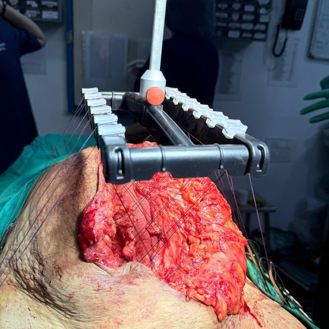
Interoperative Fascial Traction
The fasciotens® hernia device was applied for anterior fascial traction, allowing sucessful low-tension closure of the anterior rectus sheath.
%20-%20Open%20Approach%2015cm_photo10.jpg?width=3120&height=4160&name=Mahadar%20(Mumbai)%20-%20Open%20Approach%2015cm_photo10.jpg)
Skin Closure
The subcutaneous tissue was closed in layers and skin stapled. The patient was extubated on the table and had an uncomplicated recovery. She was mobilized on postop day 1 and discharged home on postop day 7.
---Open-Approach-Post-op-CT.gif?width=540&height=484&name=Mahadar-(Mumbai)---Open-Approach-Post-op-CT.gif)
Postoperative CT Scan
Follow-up imaging after reconstruction. The abdominal contents are restored to their proper position with reestablished midline continuity, showing effective defect closure and restoration of domain.
Start with fasciotens yourself
