A 22-year-old patient with a notable medical history of exploratory laparotomy for perforated appendicitis performed four years ago, which was followed by a stormy post-operative course. The patient subsequently developed a hernia at the previous surgical site. Comorbid conditions include poorly controlled diabetes mellitus (initial HbA1c: 11.2 %) and morbid obesity (BMI: 37 at first consultation). Lifestyle factors include chronic smoking and regular alcohol use.
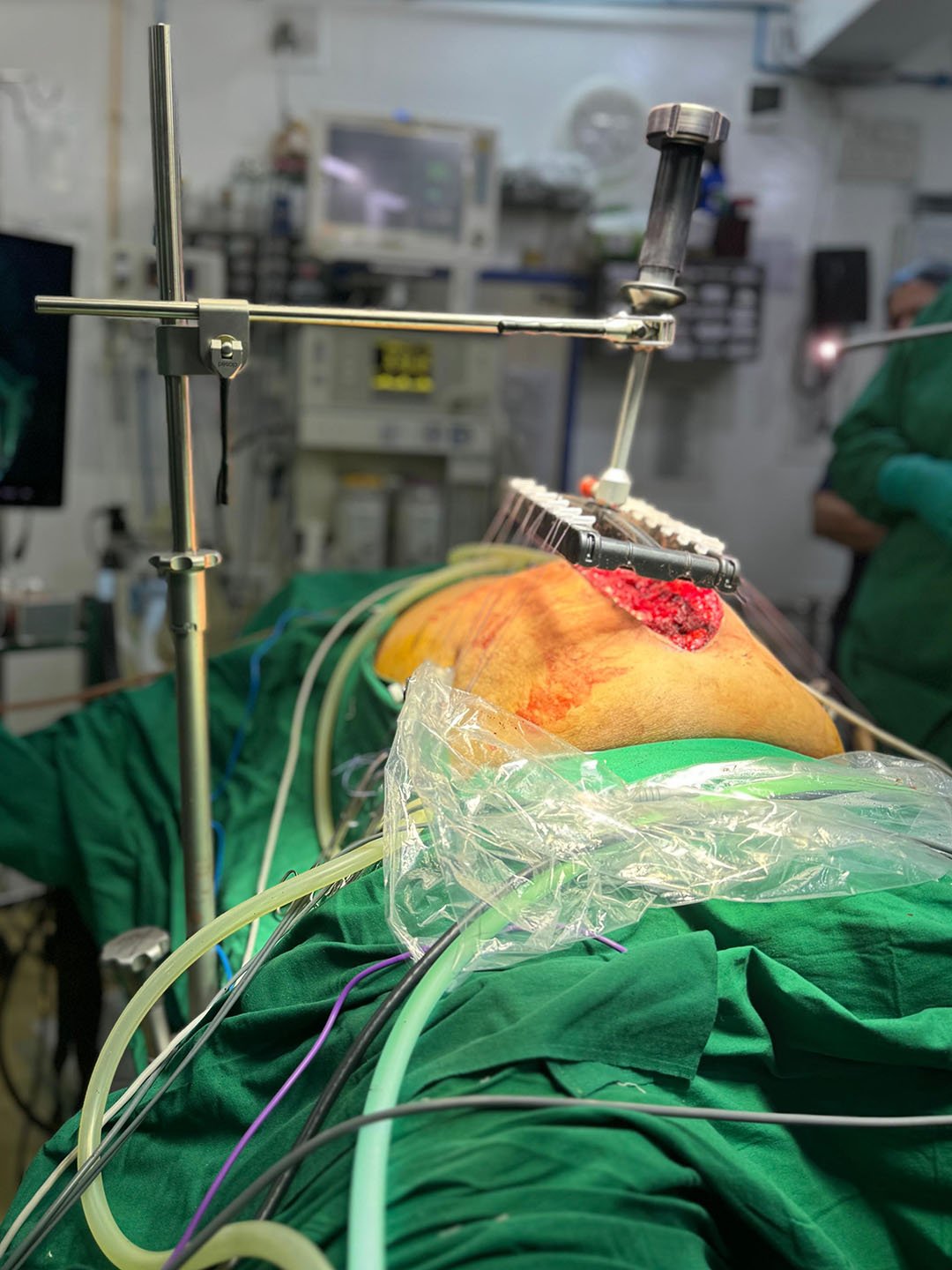
IFT in a hybrid approach
Hybrid Approach in a Large Incisional Hernia
Jeevanshree Hospital, Maharashtra, India – July 2024
Case Overview
- Patient History
- Preoperative Notes
- Surgical Plan
- Intraoperative Details
- Postoperative Course
Despite counseling on the need for strict glycemic control and weight reduction, the patient returned one year later with worsening clinical parameters: an increased BMI of 39.4 (weight: 118 kg) and an HbA1c of 9.8 %.
Preoperative Assessment:
Preoperative investigations revealed a large right paramedian hernia defect measuring 15 x 22 cm. Pulmonary function tests indicated mild obstructive airway disease. Upper gastrointestinal endoscopy (OGD scopy) showed normal findings, and the patient was cleared for surgery from a cardiac standpoint.
The patient was initial advised to undergo a two-stage approach but insisted on a single combine surgery due to psychological distress. Given the complexity of the case, a single surgery staged hybrid surgical approach was planned:
Stage 1 – Laparoscopic Sleeve Gastrectomy:
This was performed as the initial step to promote metabolic improvement and facilitate weight reduction prior to definitive hernia repair.
Stage 2 – Hybrid Hernia Repair:
This involved a laparoscopic Rives-Stoppa dissection combined with a right-sided Transversus Abdominis Release (TAR). The entire hernia sac was preserved to enable closure of the posterior rectus sheath. fasciotens®Hernia was utilized to aid in the approximation of the large anterior fascial defect. Additionally, open revision of the previous surgical scar was performed.
Initial laparoscopic gastric sleeve was performed. Followed by eTEP, open unilateral Transversus Abdominis Release (TAR) and posterior rectus sheath closure using the preserved hernia sac. The residual anterior fascial defect measured 12 cm. Use of fasciotens®Hernia enabled gradual tension reduction and successful approximation of the anterior fascial edges, allowing for primary closure of the defect.
Duration: Total operative time was 7 hours and 25 minutes.
Anesthesia Management: Peak and plateau airway pressures remained well controlled throughout the procedure, indicating stable intraoperative respiratory parameters.
The patient was extubated on the operating table and demonstrated a smooth initial recovery. Ambulation began the following morning in the ward, and the patient was discharged on the 4th postoperative day in stable condition.
At the 5-month follow-up, the patient showed significant clinical improvement. Weight had reduced to 79 kg (BMI: 26.4), and diabetes was well-controlled with an HbA1c of 6.9. There was no evidence of hernia recurrence, and the patient reported excellent functional and cosmetic outcomes.
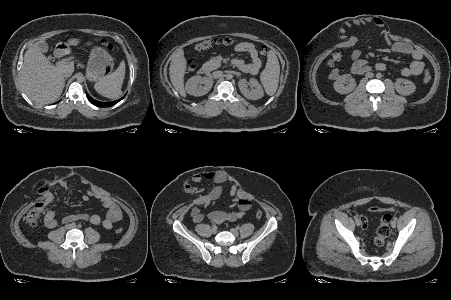
Pre-operative CT Scan
The CT scan shows ventral hernia mainly in the middle and lower part of the abdomen. The hernia sac contains small bowel loops, the rectus muscles and lateral abdominal wall appear intact.
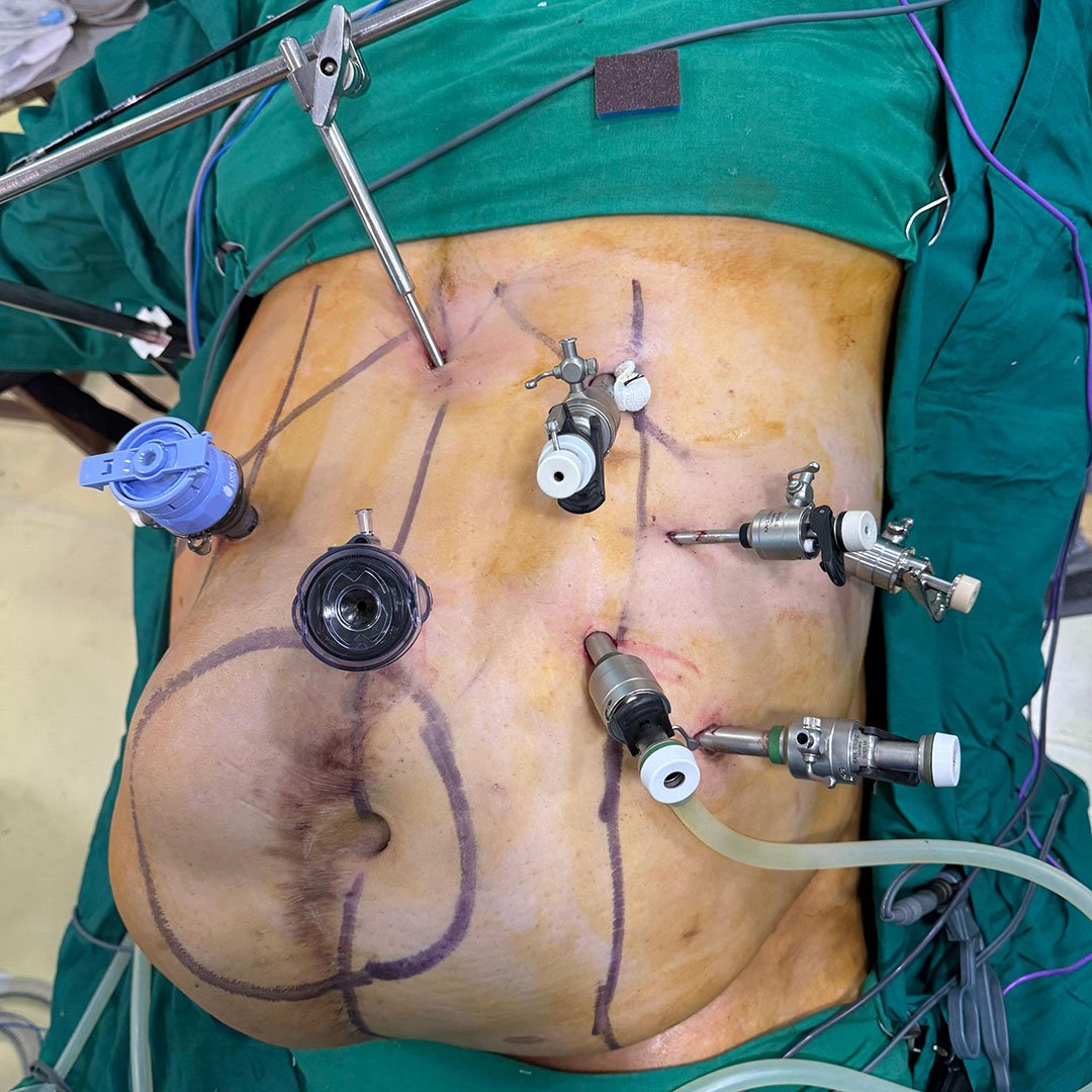
Trocar Placement and Initial Laparoscopic Access
A laparoscopic approach was initiated with multiple ports placed strategically to perform sleeve gastrectomy as well as eTEP dissection. The large right paramedian hernia was mapped on the abdominal wall.
%20Hybrid%20Intraop%204.jpg?width=1600&height=1156&name=Mahadar%20(Mumbai)%20Hybrid%20Intraop%204.jpg)
Laparoscopic Gastric Sleeve
Following trocar placement, a laparoscopic sleeve gastrectomy was performed as the first stage of this combined procedure.
%20Hybrid%20Intraop%201.jpg?width=4032&height=2989&name=Mahadar%20(Mumbai)%20Hybrid%20Intraop%201.jpg)
eTEP
Retrorectus dissection carried out using the eTEP approach, allowing wide exposure of the hernia defect margins and adequate mobilization of the posterior sheath for layered closure.
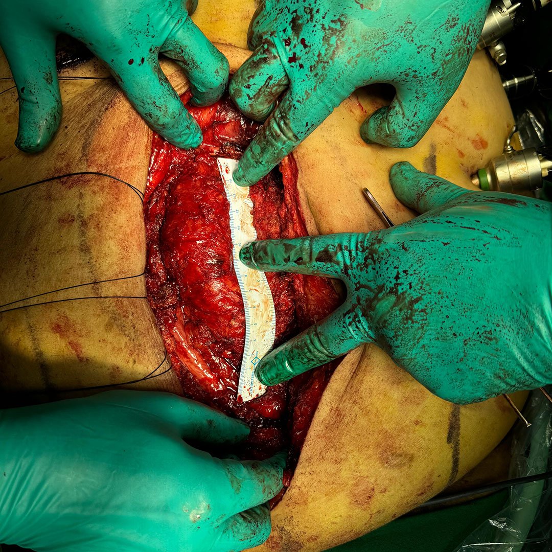
Open Surgery
Open exposure of the hernia sac with posterior dissection and measurement. The hernia sac was initially and the posterior rectus sheath was eventually reconstructed using a peritoneal flap, aided by a right-sided Transversus Abdominis Release (TAR). A mesh was placed in sublay position.
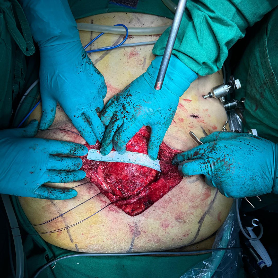
Defect Measurement and Preparation for IFT
At this point, the anterior transverse fascial defect measured approximately 12 cm. The decision was made to apply fasciotens®Hernia to facilitate anterior sheath closure without creating an increase in the intra-abdominal pressure.

IFT Using fasciotens®Hernia
fasciotens®Hernia was mounted and used to apply continuous traction to anterior rectus sheaths. This significantly reduced the fascial gap, enabling complete closure of the anterior layer without bridging.
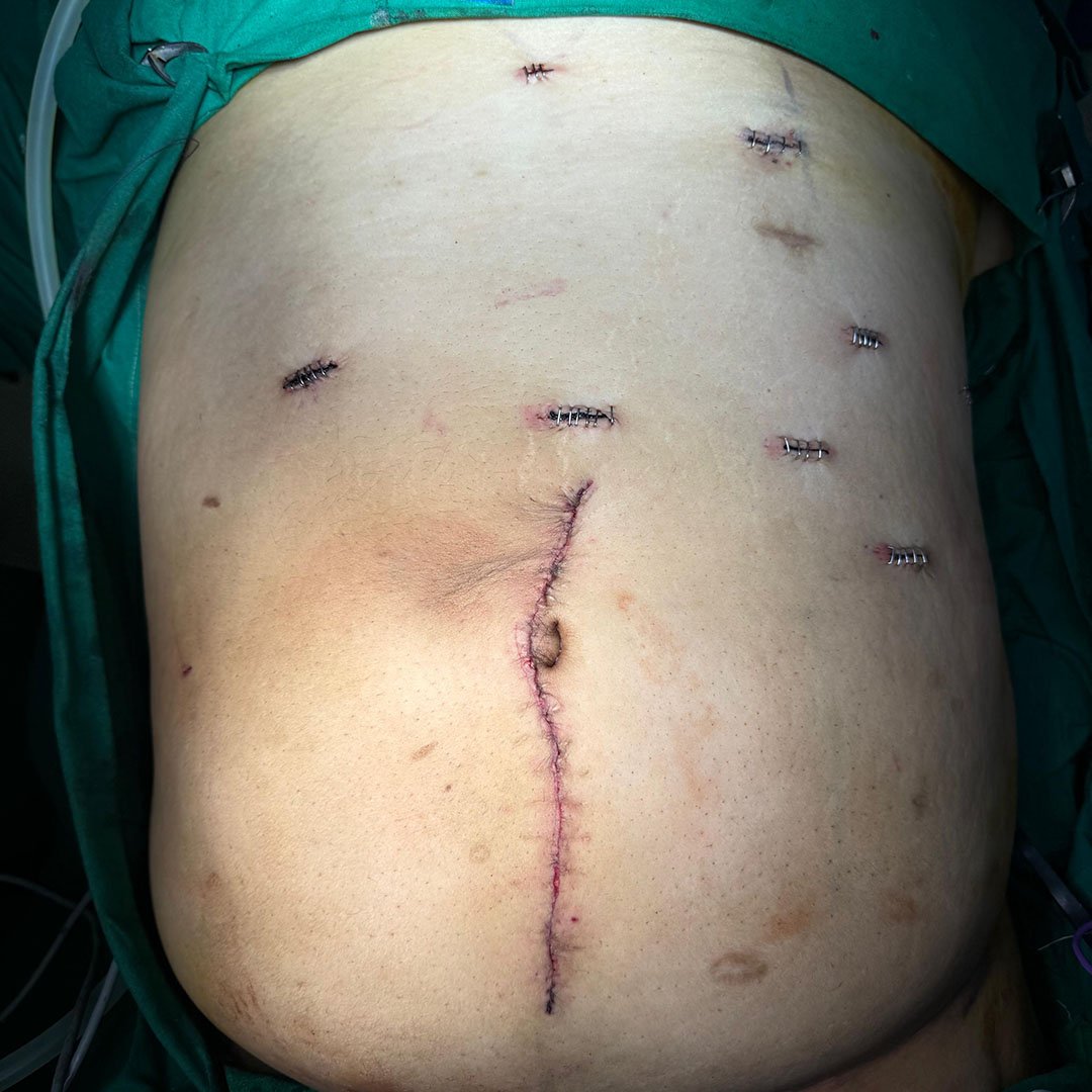
Fascial Closure and Wound Management
Immediate postoperative view with closure and stapled port sites. The fascia was fully closed, and the anterior sheet approximated without the need of bridging. No increase in ventilation pressures was noted, and the patient remained hemodynamically stable throughout. The patient was extubated on the table and experienced an uneventful recovery. He was mobilized early and discharged on postoperative day 4.
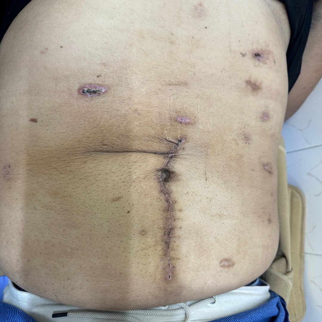
5-months Follow-up
At the 5-months follow-up, his BMI had reduced to 26.4, and his HbA1c was well-controlled at 6.9 %. There was no evidence of a hernia recurrence.
Functional Result
Watch the video on the right to observe the abdominal wall under dynamic strain (during patient coughing), demonstrating intact structure, no bulging, and favorable healing following the combined procedure.Start with fasciotens yourself
