
Complex Hernia with Infected Mesh
IFT in a case of multi-recurrent incisional hernia with exposed, infected mesh
Cisanello University Hospital, Pisa, Italy - February 2025
Dr. Luigi Cobuccio reports this case involving a 56-year-old male with a history of multiple failed abdominal wall reconstructions, presenting with exposed mesh, chronic infection, and recurrent hernia. The clinical scenario was further complicated by previous mesh removals, surgical site infection, and a lack of prehabilitation options. Given the challenging surgical field and patient factors, an open approach was planned with the use of intraoperative fascial traction to facilitate direct closure.
Case Overview
- Patient History
- Preoperative Notes
- Surgical Procedure
- Postoperative Course
A 56-year-old obese man (BMI 36.5), ex-smoker, presented with a complex history of recurrent umbilical incisional hernia and multiple failed repairs performed at other institutions. He had previously undergone umbilical hernia repair with a preperitoneal mesh in 2022, followed by laparoscopic cholecystectomy and suture hernia repair in 2023, which was complicated by small bowel obstruction, re-exploration, and mesh removal. In 2024, he underwent an open Rives-Stoppa repair with polypropylene mesh and anterior component separation (M2M3 W3 defect), followed by a significant postoperative retromuscular hematoma requiring bilateral angioembolization. The course was complicated by surgical site infection and dehiscence, ultimately exposing the mesh.
The patient presented with exposed mesh (4 cm diameter), surrounding erythema, and evidence of hernia recurrence (13 cm wide on CT) cranially associated with rectus diastasis. Wound swabs showed Staphylococcus aureus and Corynebacterium simulans. Due to the progressively enlarging exposed mesh and poor compliance, it was decided to proceed with surgical intervention. Prehabilitation was not feasible (no botulinum toxin A or weight loss possible).
Surgical exploration revealed significant inflammation and a 15 cm wide residual fascial gap. Complete mesh removal was performed en bloc with erythematous skin. Fasciotens®Hernia was applied to the anterior and posterior rectus sheaths (where present) for 28 minutes to facilitate medialization. This allowed for tension-free midline fascial closure using small-bite PDS 0 sutures. One subcutaneous drain was placed for seroma prevention.
Surgical Steps

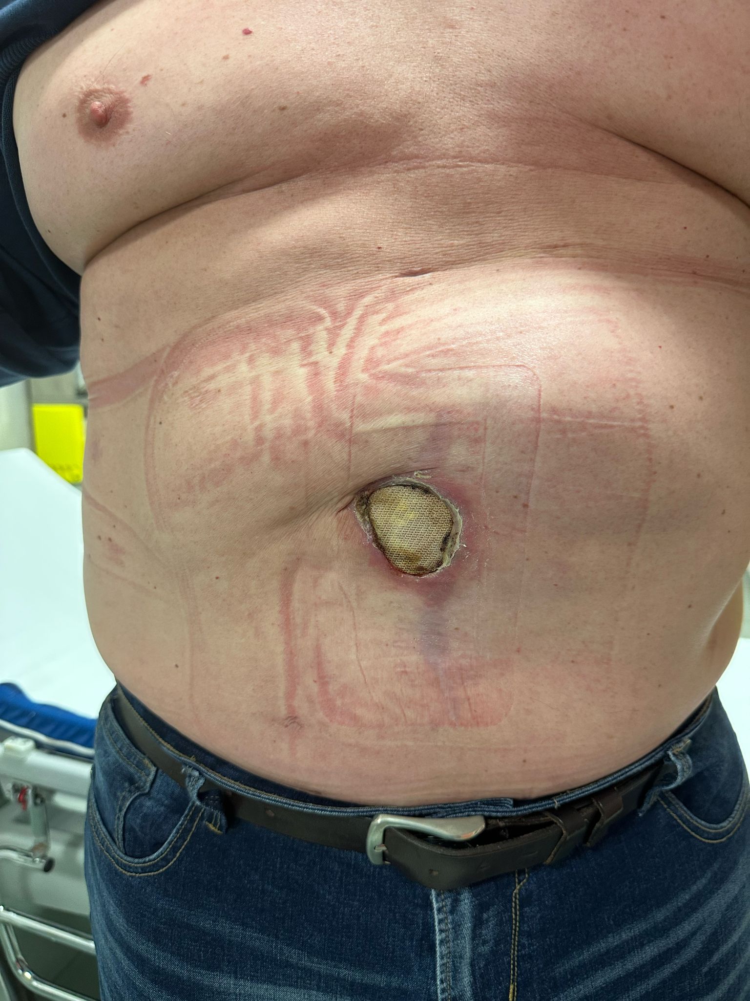
Clinical Presentation
Initial presentation showing exposed mesh at the umbilicus with surrounding erythema.
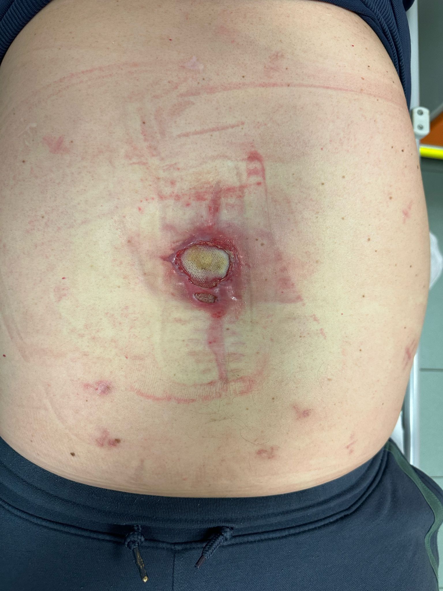
Standing Profile
Standing view illustrates the extent of abdominal wall involvement and the hernia bulge.

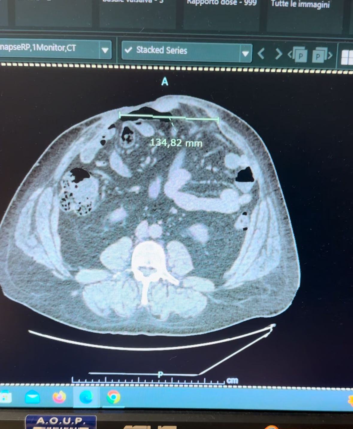
Pre-operative CT Scan
13.4 cm hernia recurrence with cranial rectus diastasis.
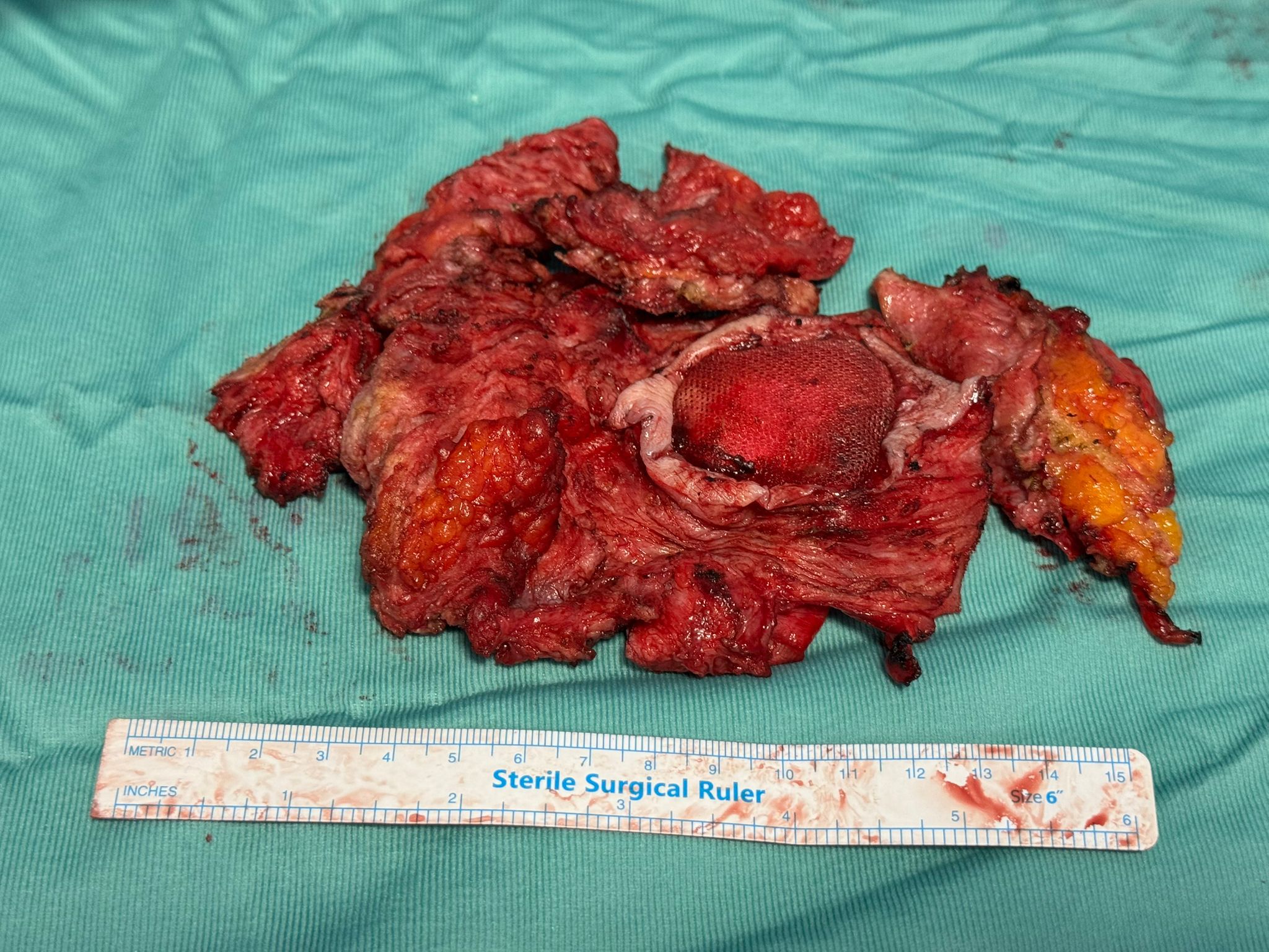
Mesh Explantation
Infected mesh removed en bloc with inflamed overlying skin.
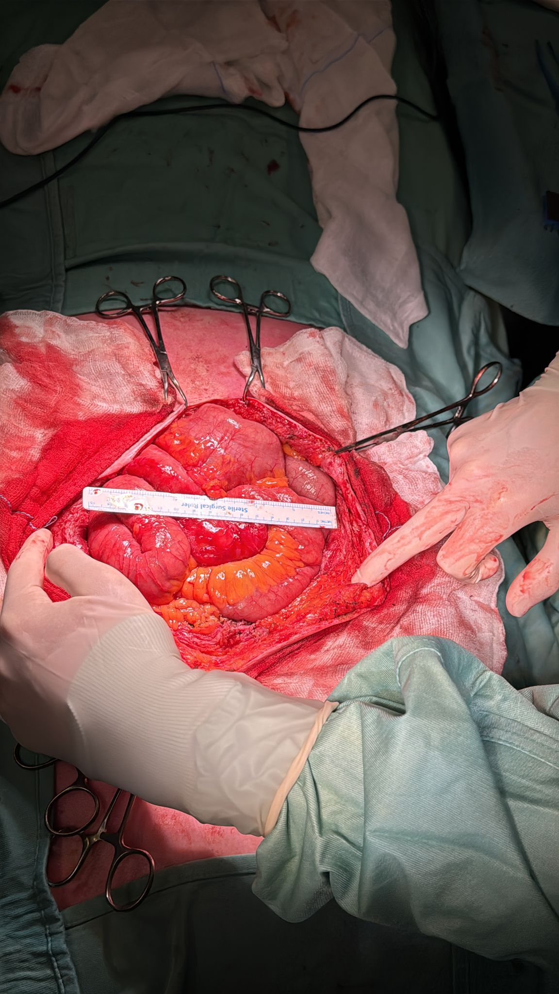
Fascial Defect
15 cm midline gap identified intraoperatively.
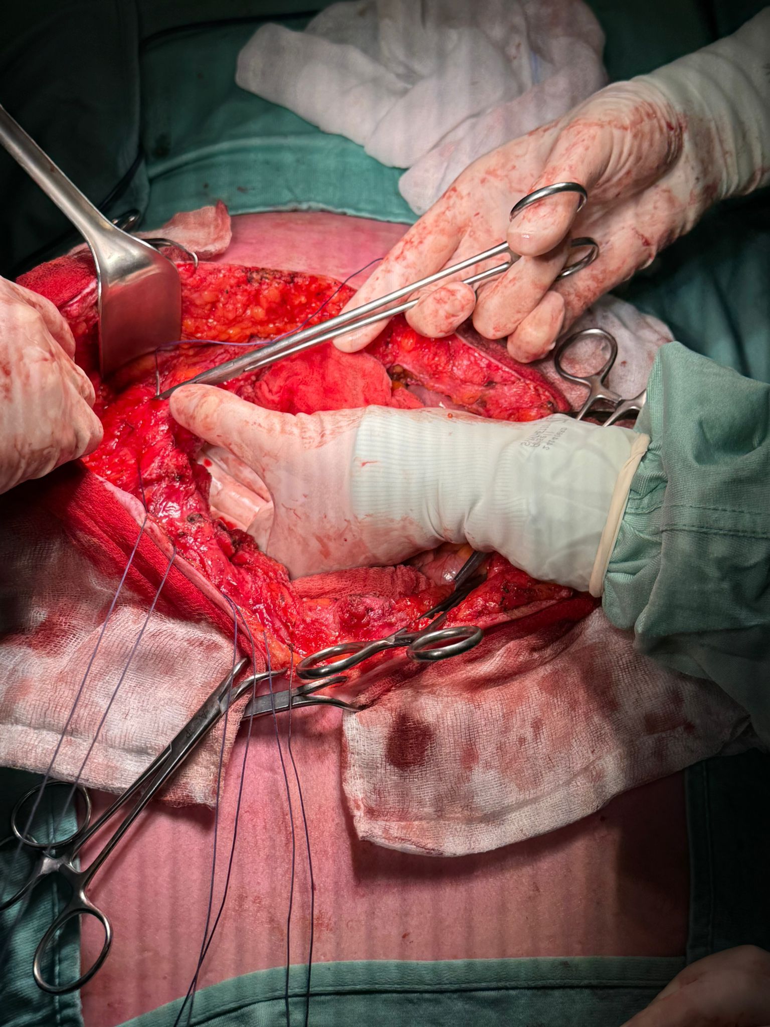
Suture Setup
Sutures placed along fascial edges prior to traction.
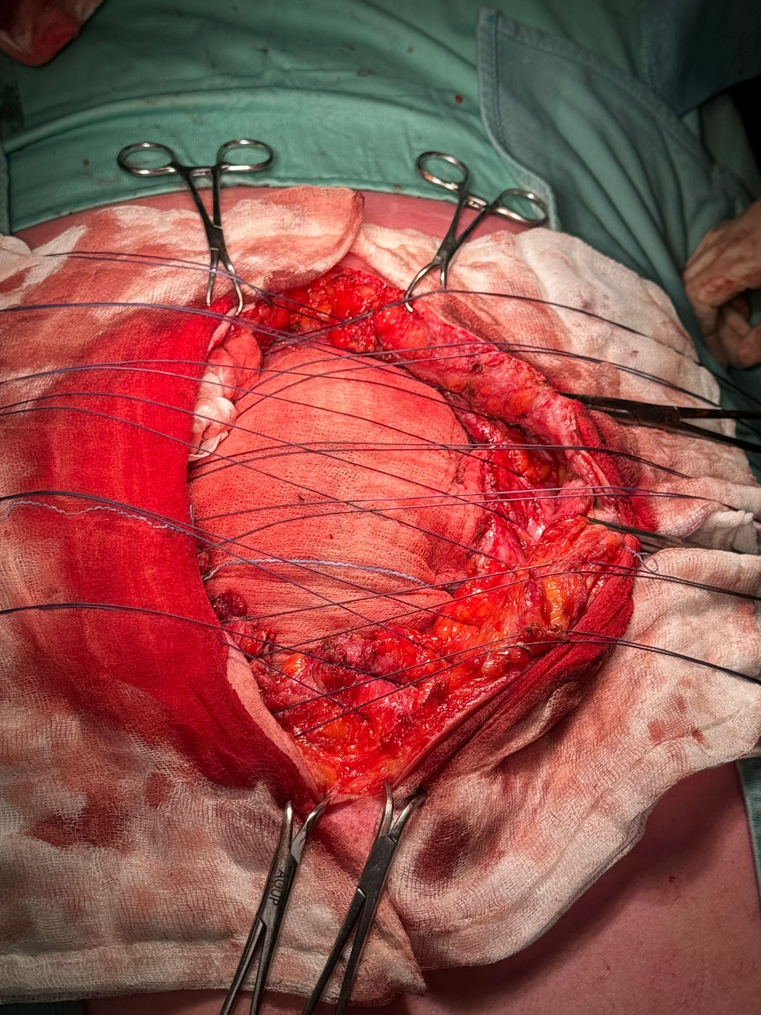
Ready for Traction
Sutures arranged for attachment to fasciotens®Hernia.
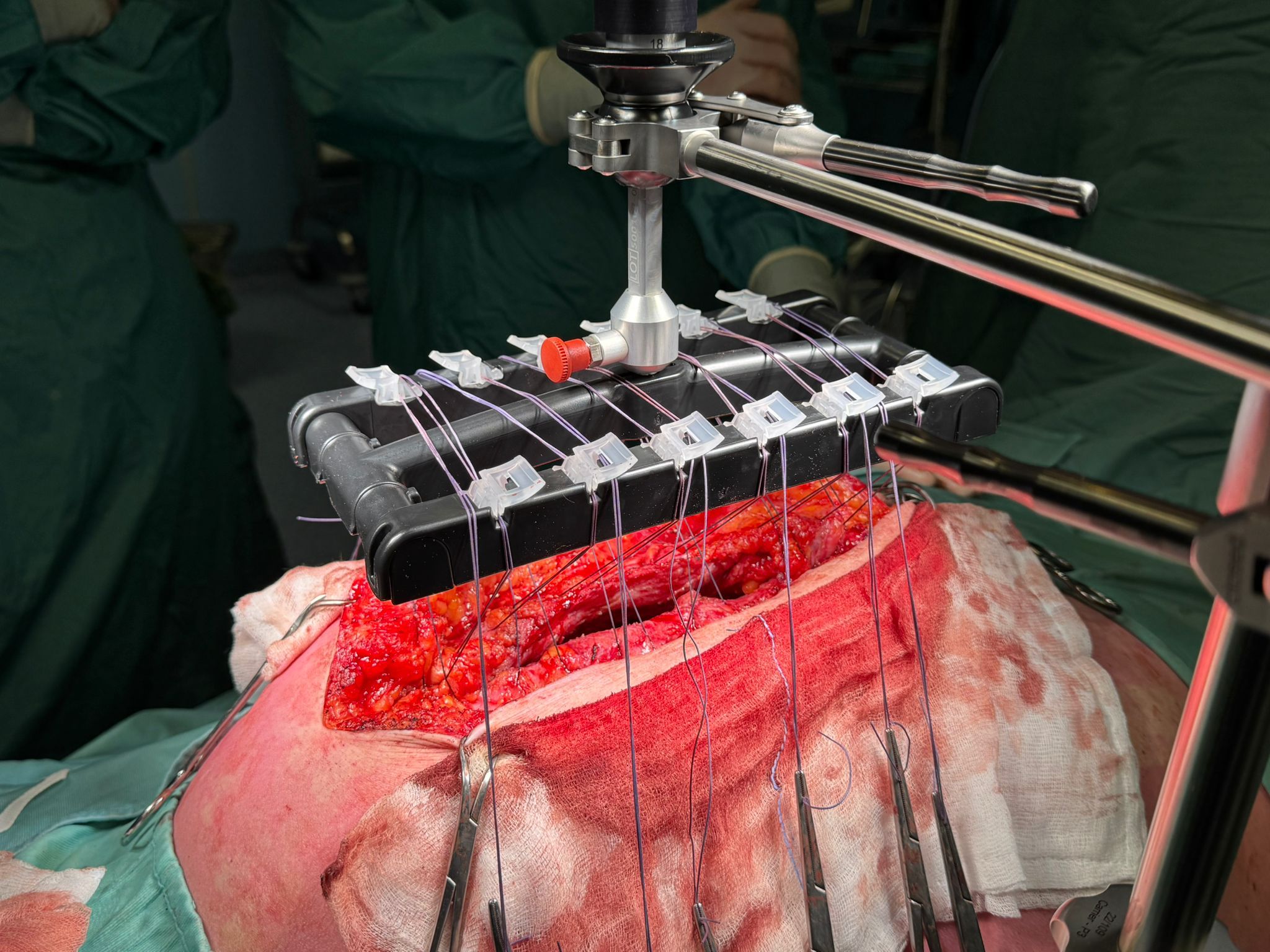
Sutures Secured
Traction applied using fasciotens®Hernia system to elongate abdominal wall
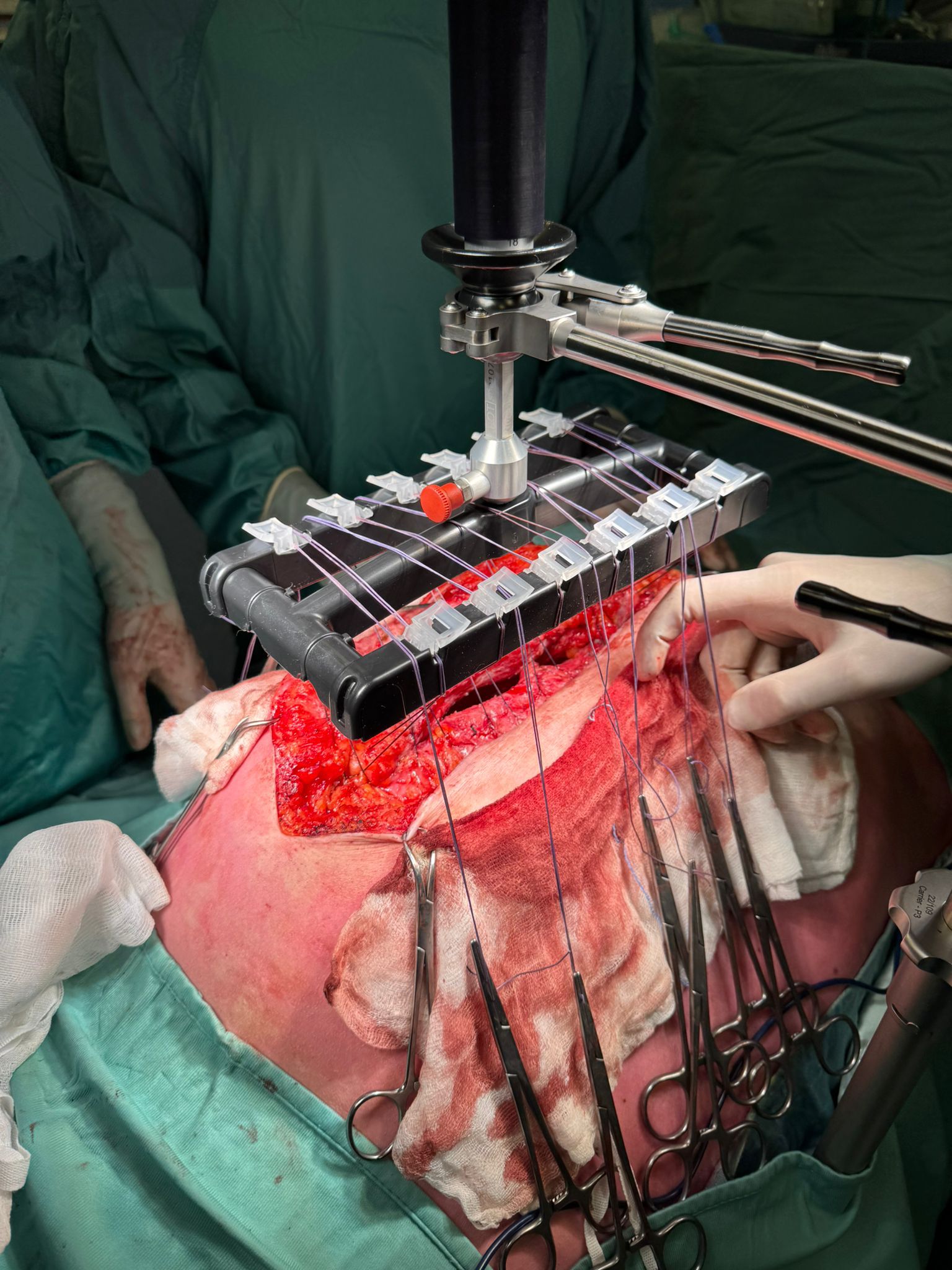
Active Traction
Controlled, dynamic vertical traction applied for 28 minutes.

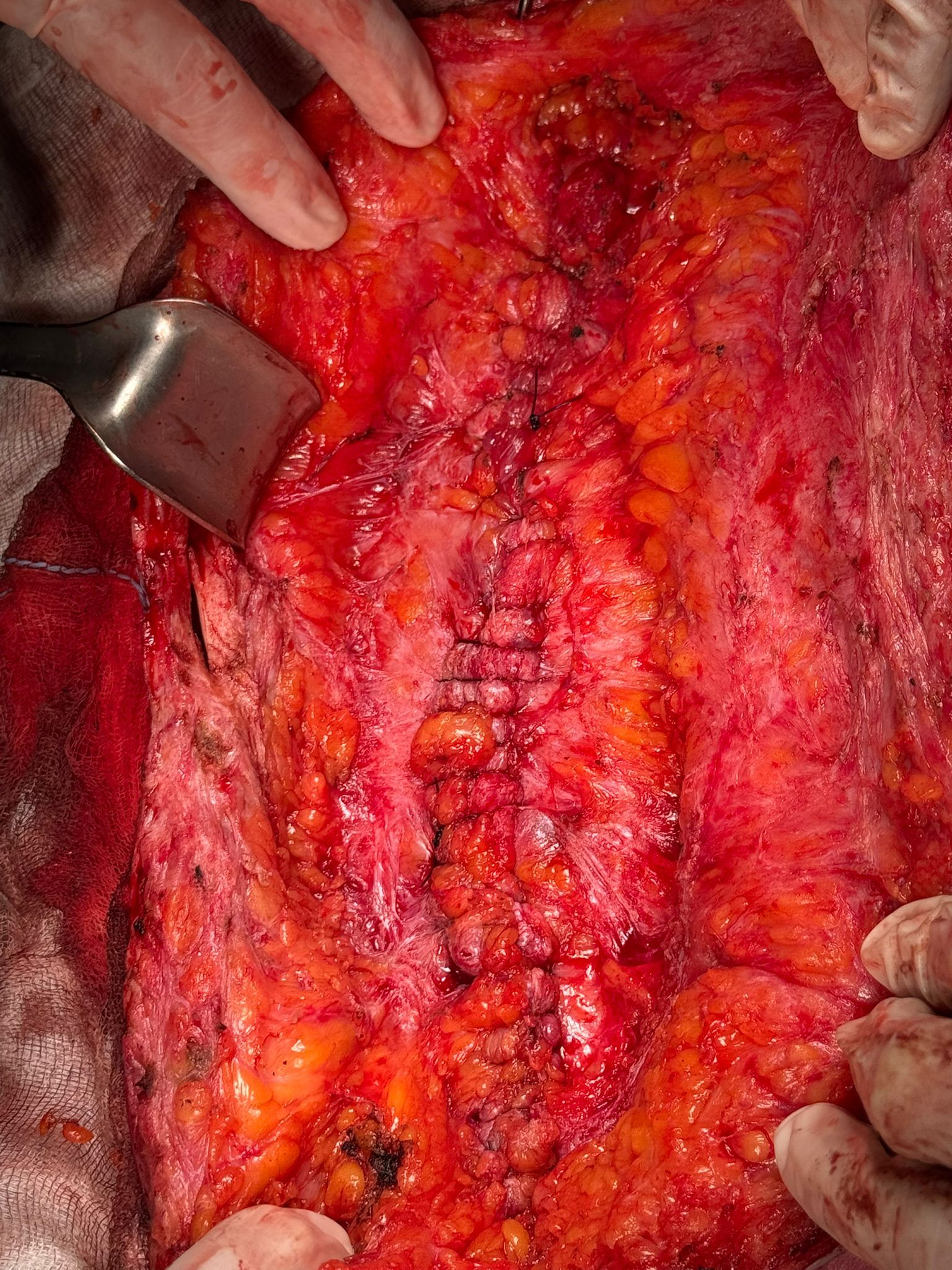
Direct Fascial Closure
Midline successfully re-approximated after IFT.

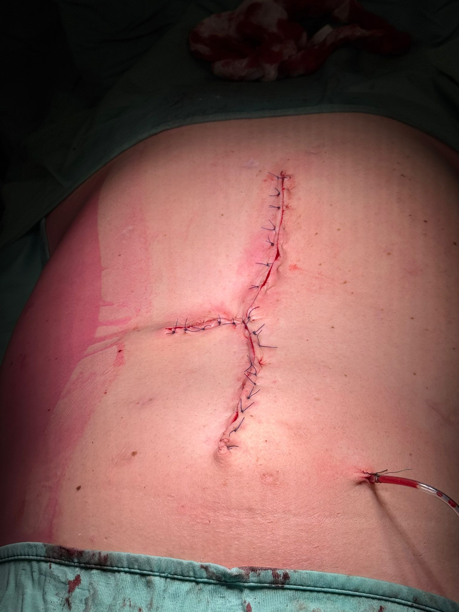
Final Interoperative View
Completed closure of abdominal wall and skin.
Start with fasciotens yourself